


Image Sequence
Existing X-Ray fluoroscopy hardware is used to capture cinematic image sequences of the patient’s lungs under ventilation from five different views.

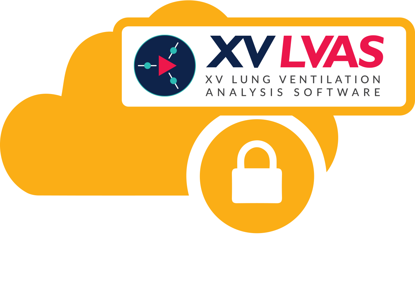
4DMedical XV LVAS
Utilizing military-grade security technology, 4DMedical conducts the analysis and returns a report that can be accessed by both the clinician and the patient using the XV LVAS platform.
XV LVAS Ventilation Report
A color-coded visualization is generated, showing both coronal and axial slices at peak inspiration, plus a 4-dimensional animation.
The report also quantifies ventilation heterogeneity, which is a widely recognized indicator of lung health.
Laurel Bridge (LB)
- Fluoroscopy images are delivered to LB Site Router via the PACS
- Prior CT Exams are requested and delivered via hospital PACS to LB Site Router
- Fluoroscopy & CT Exam images are de-identified by the LB Site Router and securely sent via the internet to the LB Hub router located at 4DMedical
- 4DMedical returns an XV LVAS Ventilation Report via the LB Hub & Site Routers back to the hospital, where it is then re-identified before delivery back into the PACS/RIS.